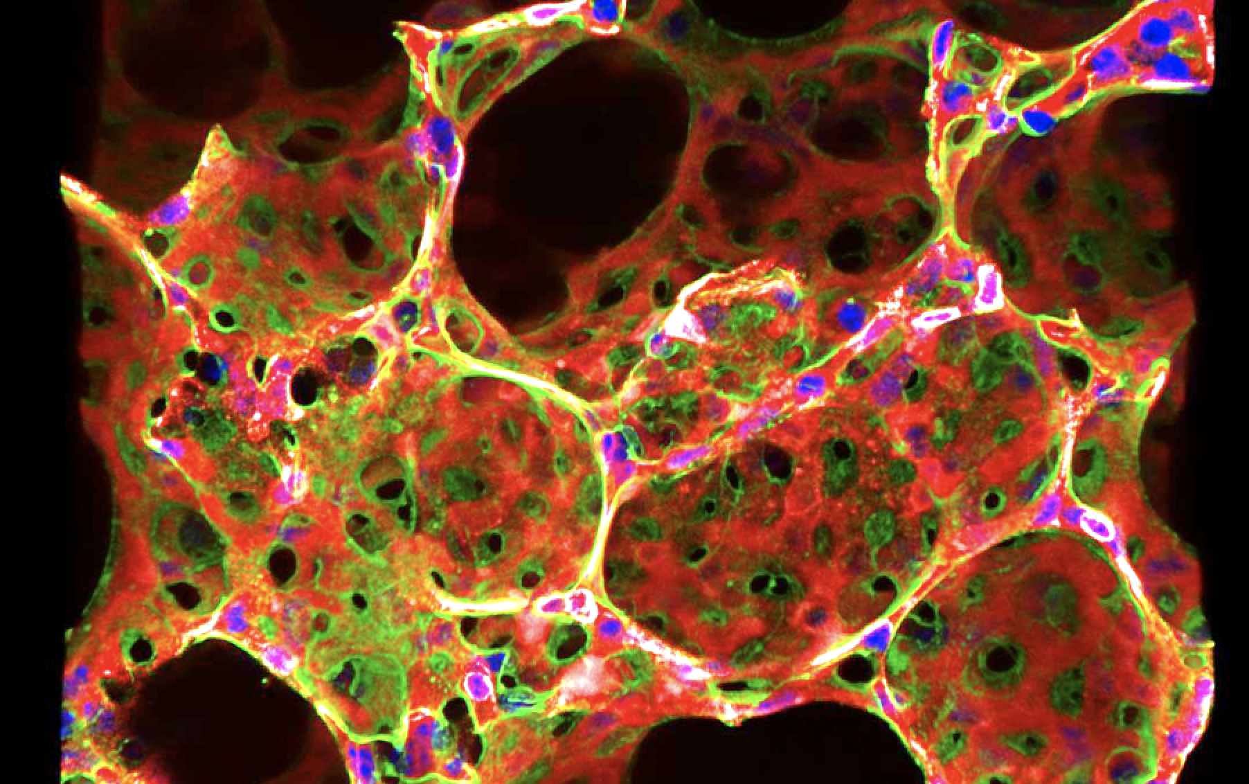
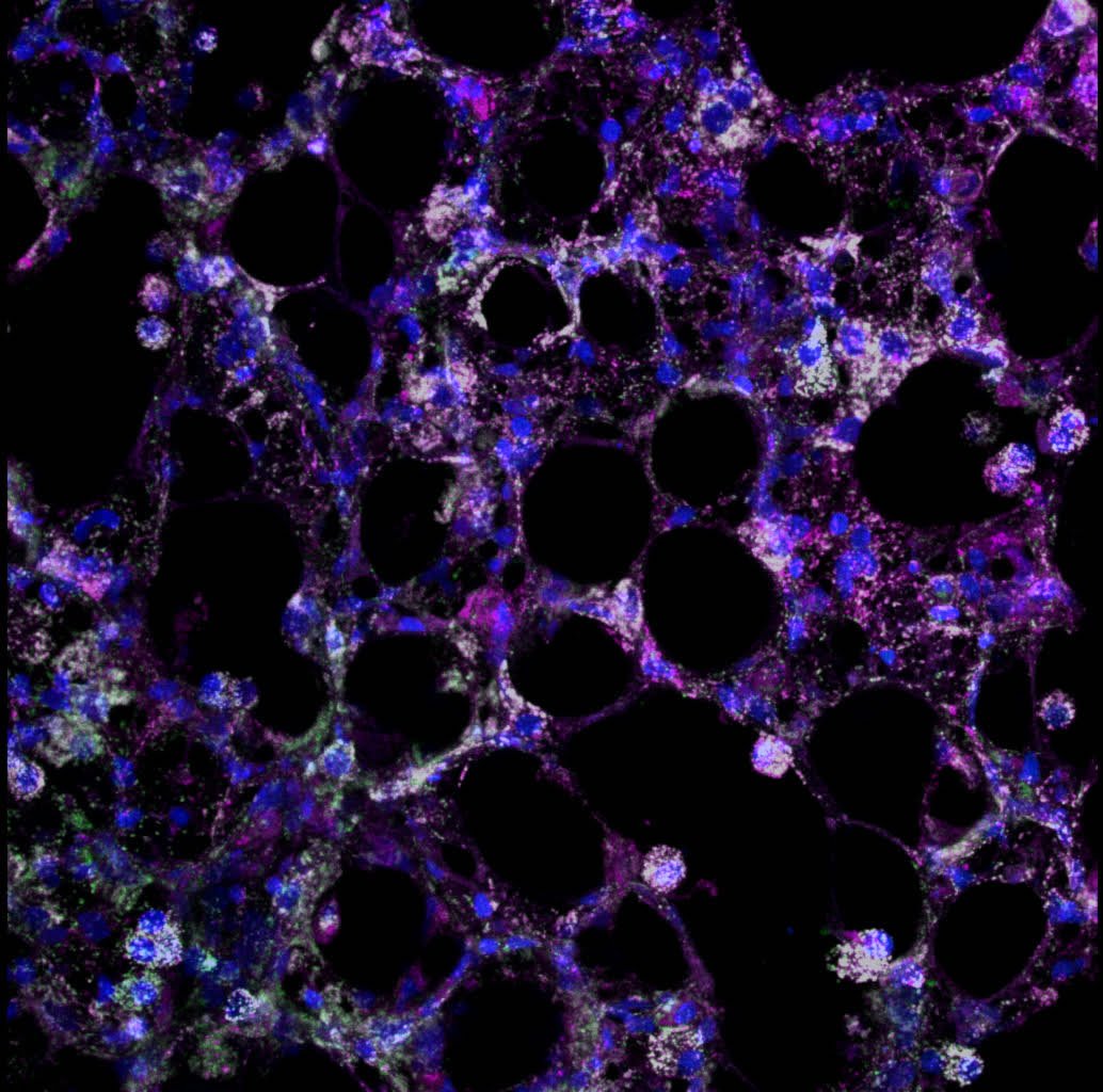
Confocal
Mouse precision-cut lung slices.
Green:Type 1 pneumocytes; Red: endothelium; Blue: DAPI
By Ke Yuan,
Yuan lab
2023 Imaging contest winner, Most Beautiful
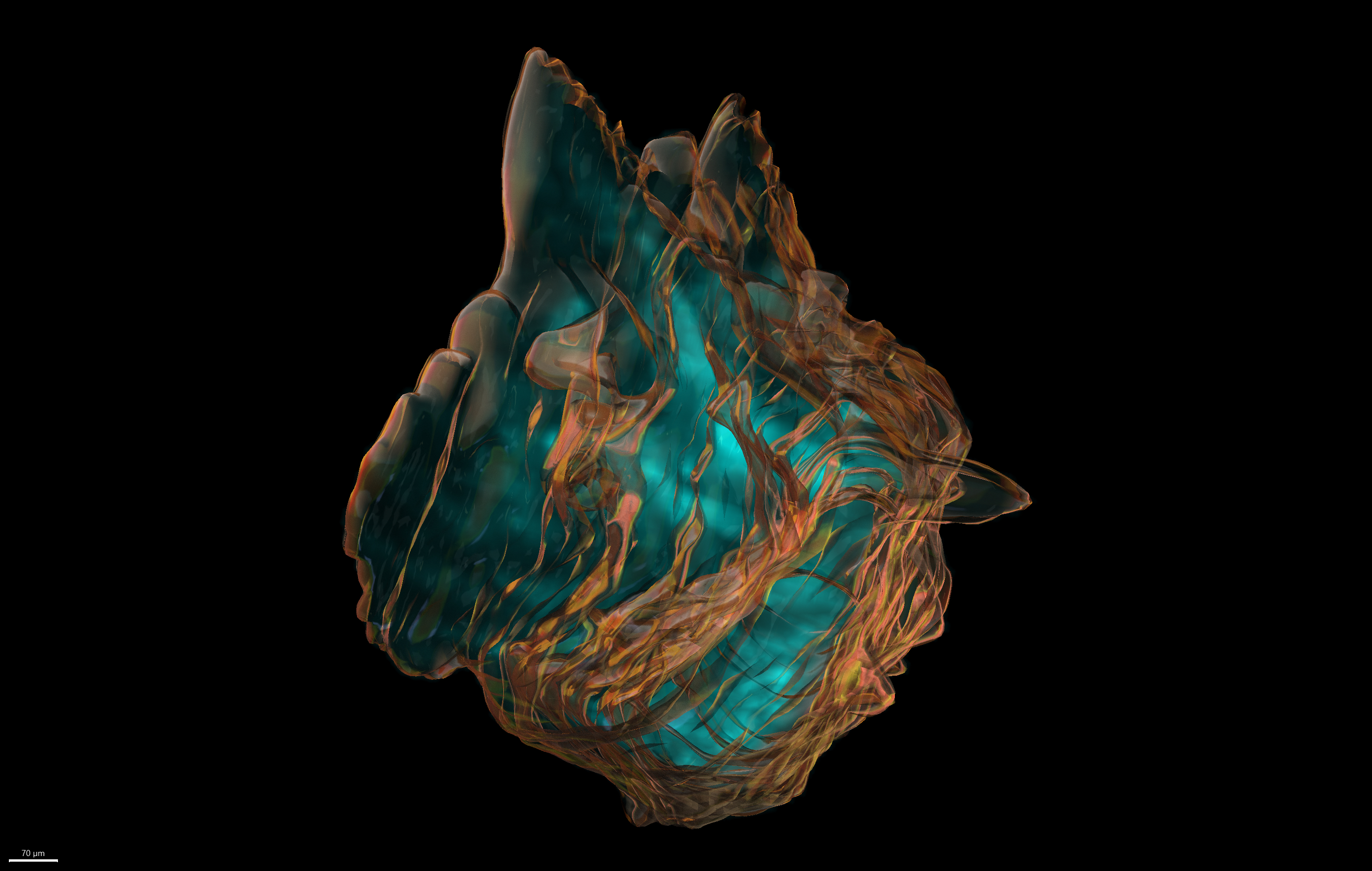
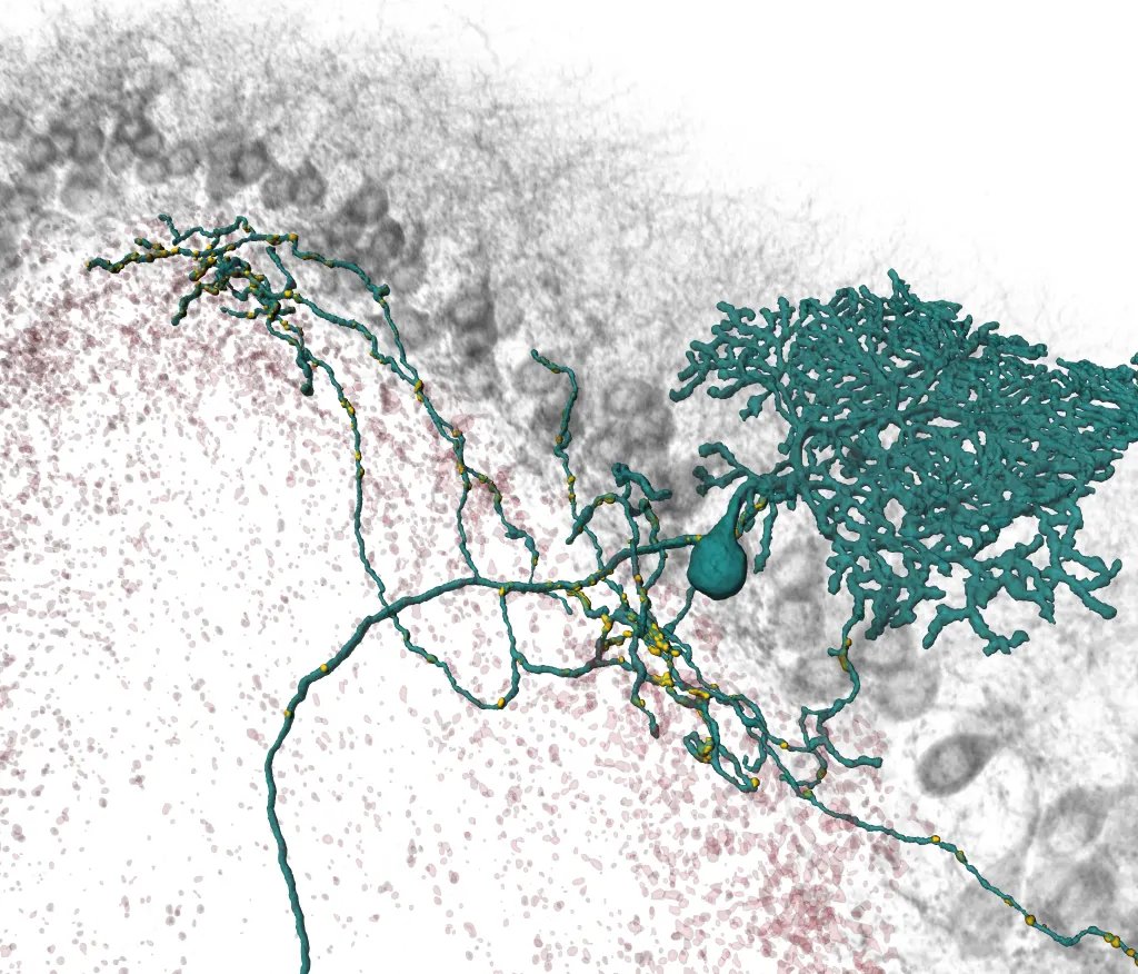
Ultramicroscope II (LaVision Biotec) light-sheet microscope; 3D reconstruction on Imaris
Atherosclerotic plaque (translucent copper surface) with CD68+ macrophages (Alexa Fluor 647 - Cyan) inside a mouse aorta. Surface was crated manually by tracing the outline of the plaque in each z-slice in the CD68-AF647 channel and then masking it before duplication. Surface texture was set to "Glass" in Imaris.
By Anurag Jamaiyar, Feinberg Lab
2023 Imaging contest winner, Most Scientific
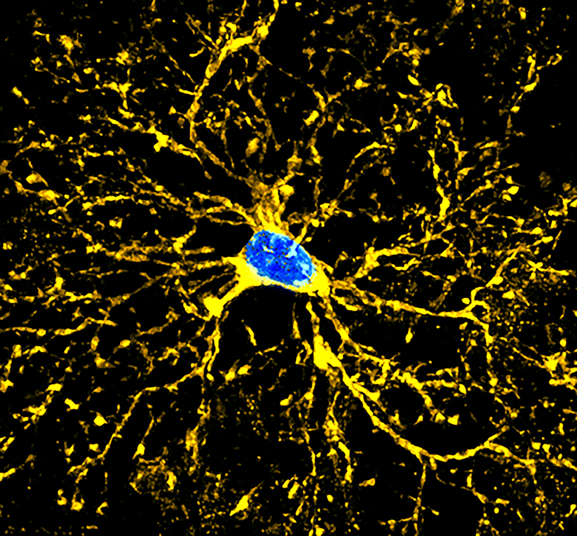
Stellaris Confocal
The Olidodendro-tree
This image shows a mature oligodendrocyte with the produced myelin. (It is taken from a transgenic mouse line where newly formed myelin can be seen through mGFP).
By Sara Conti, He Lab
2023 Imaging contest winner, Most Popular
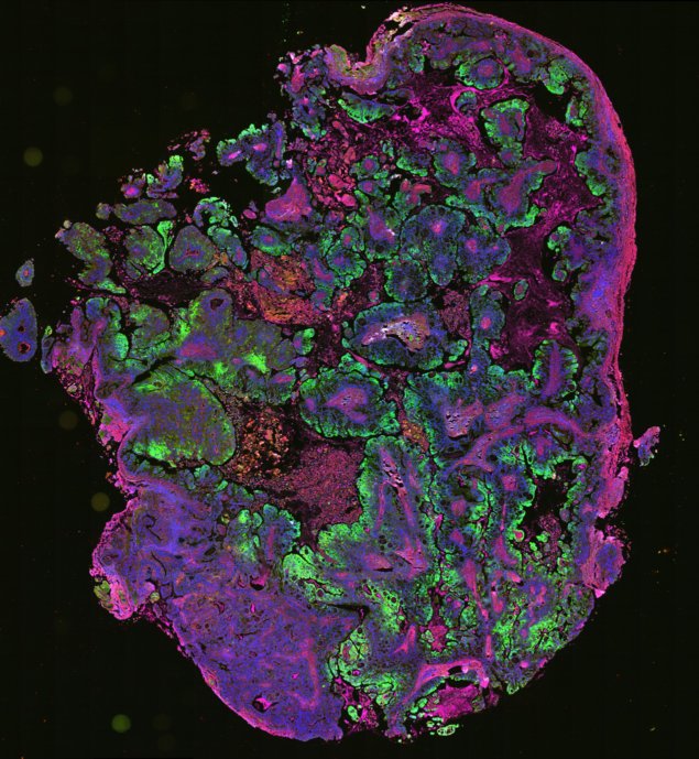
VS120 Slide Scanner
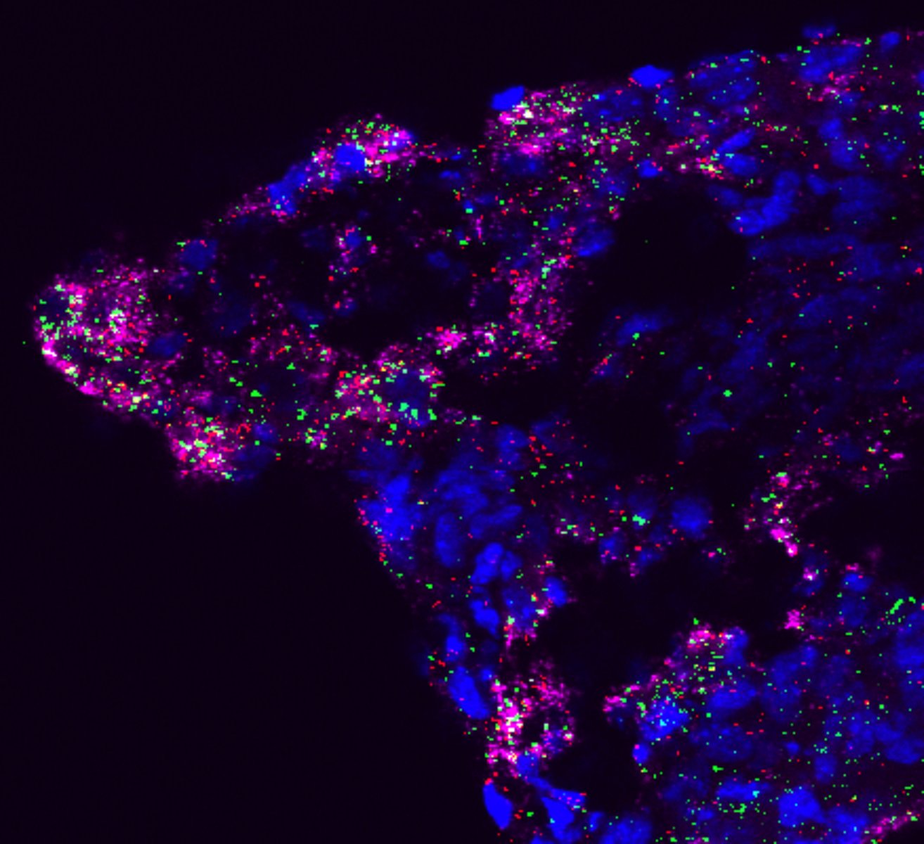
Zoomed area of the cross-section of the whole pancreatic cancer tumor xenograft stained for Collagen (Red), Fibronectin (Magenta), (representing deposition of the stroma), and phospho-NDRG1 (Green), representing activation of the NDRG1 in response to extra-cellular matrix deposition and sensing in pancreatic cancer.
By Nina Kozlova, Muranen Lab
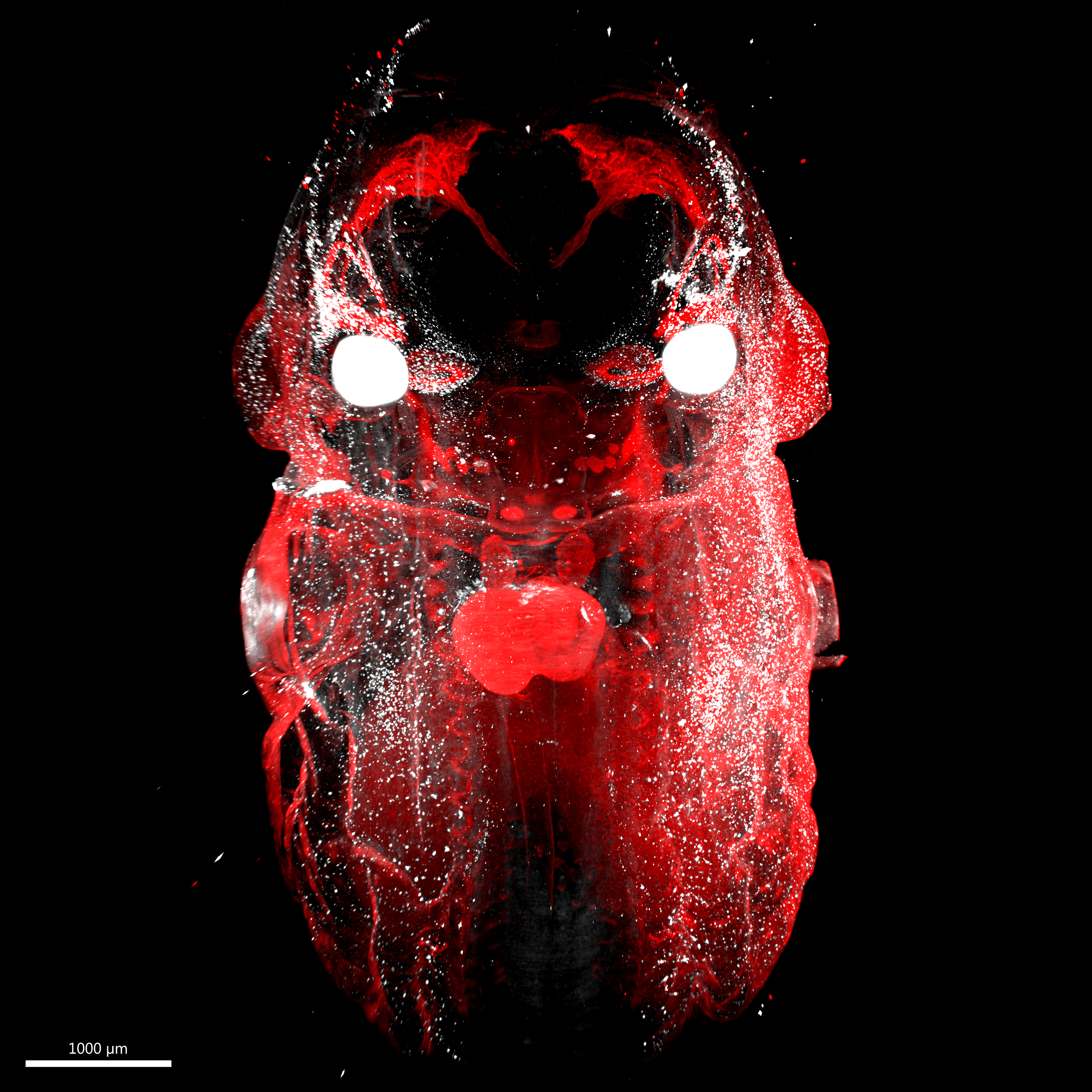
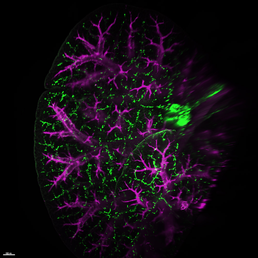
LaVision Lightsheet Microscope
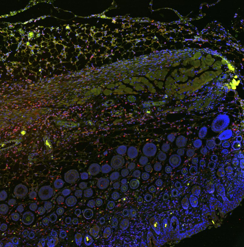
Melanoblast cells migrating through the skin from the neural crest in an e13.5 embryo
By Brian Raftrey, Hsu Lab
2022 Imaging contest winner, Most Beautiful
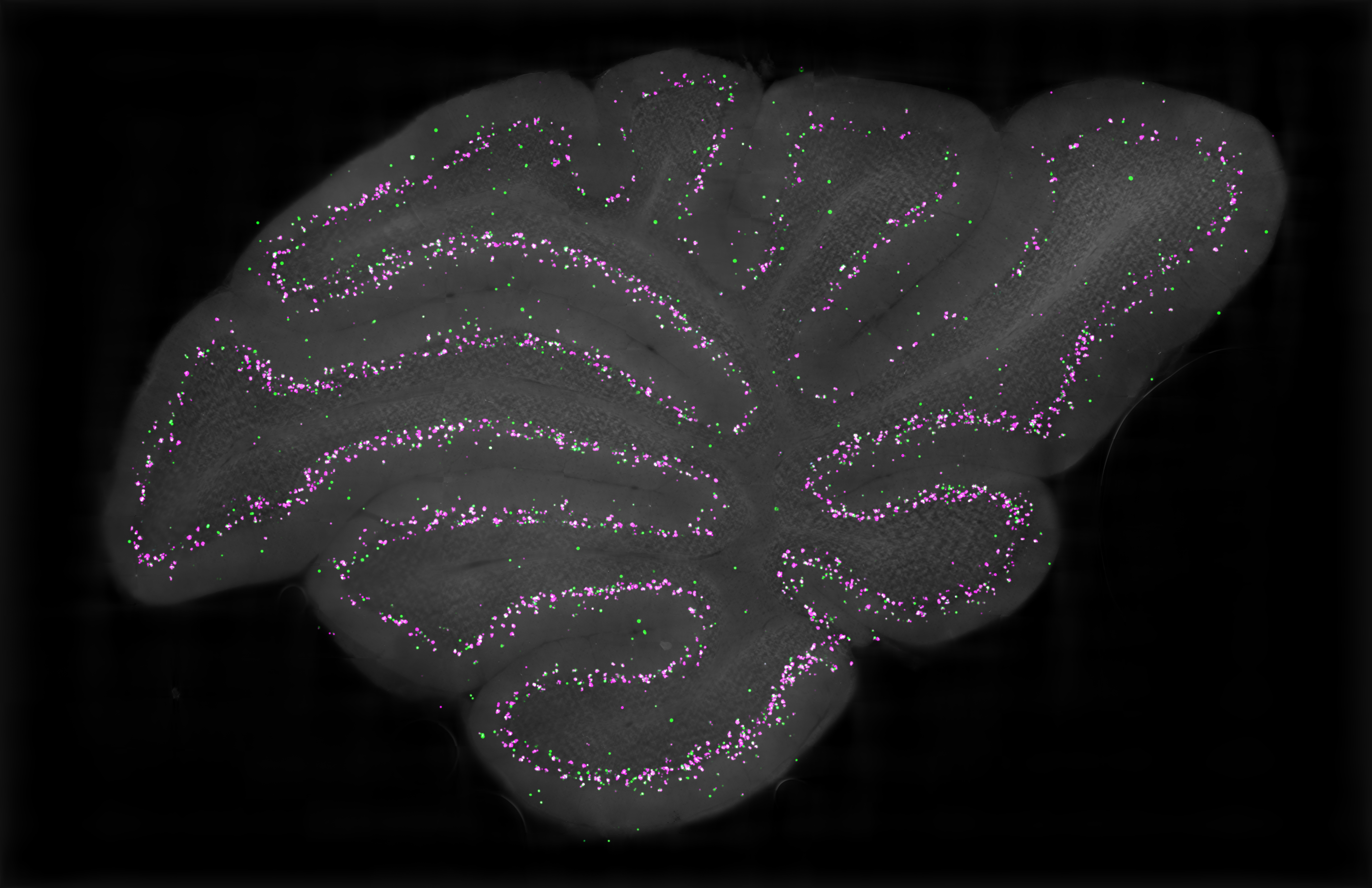
VS120 Slidescanner
Sagittal slice of cerebellar cortex showing the distribution of Candelabrum cells, a previously uncharacterized interneuron of the cerebellum.
By Tomas Osorno, Regehr Lab
2022 Imaging contest winner, Most Scientific
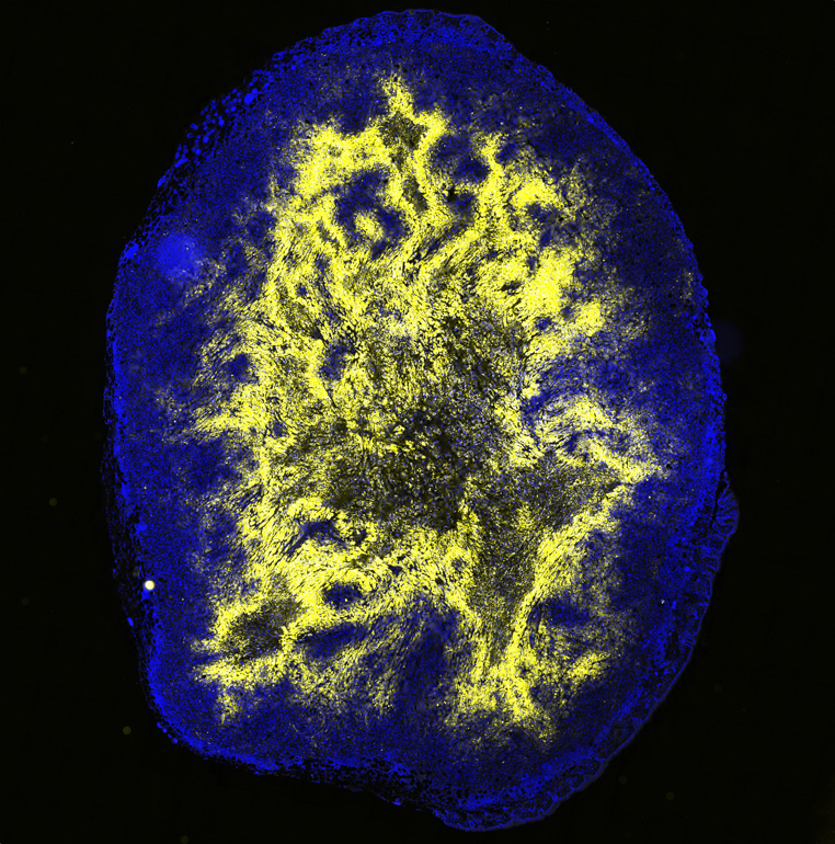
VS120 Slide Scanner
Here's a cross-section of a pancreatic tumor xenograft stained for nuclei (DAPI), target we are working on that is highly present in a hypoxic core of the tumor (RED), and a GFP-tagged inducible cassette.
By Nina Kozlova, Muranen Lab
2022 Imaging contest winner, Most Popular
Purkinjie Cells captured on Olympus FV1000 confocal rendered with Imaris (workstation 2)
RNAscope control slide or DRG captured on Olympus FV1000 confocal
Skin section captured on Olympus VS120 whole slide scanner Bertagnolli lab, BWH
Fly brain montage captures on LeicaSP8X confocal
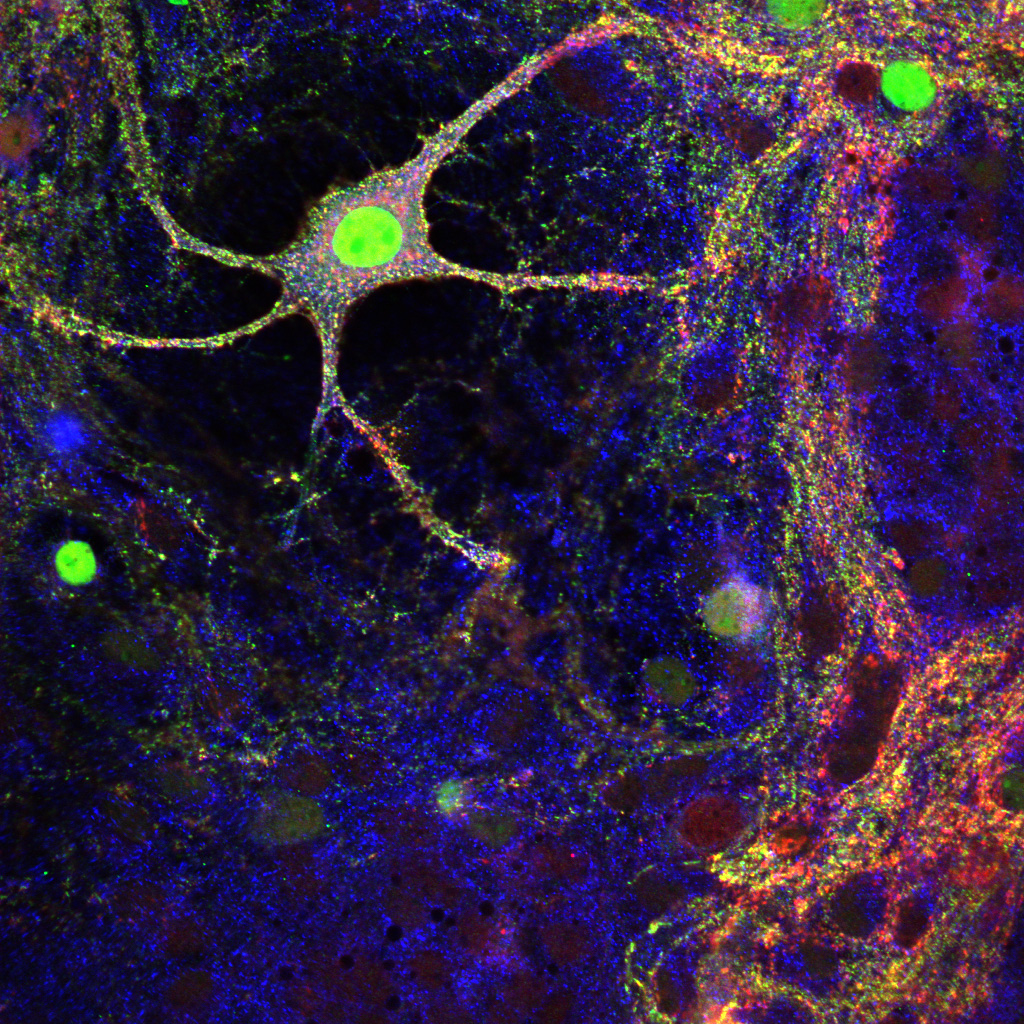
Hippocampal neuronal culture stained for three proteins: In blue: Map2, In red: Synaptophysin, in green: ELKS1. Acquired on Leica SP8X confocal. Kaeser Lab, HMS
Lung stained with Alpha smooth muscle (purple) and lymphatics (green). Acquired on laVision Lighsheet. Fine Lab, Boston University
Mouse ear stained for immune cells (green) and vasculature(fire) captured on Olympus VS120 slide scanner. Chenghua Gu Lab, HMS
Lung tissue captured on Leica SP8X stained with Hoechst (blue) Cytochrome c (green) and mitotracker (red). Kristopher Sarosiek lab, HSPH
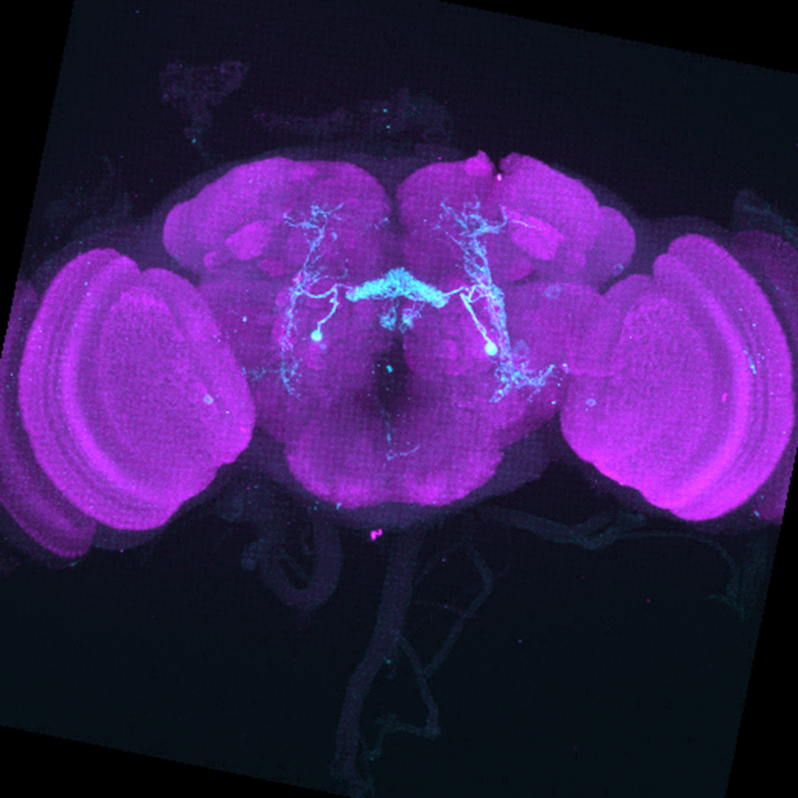
Leica SPE
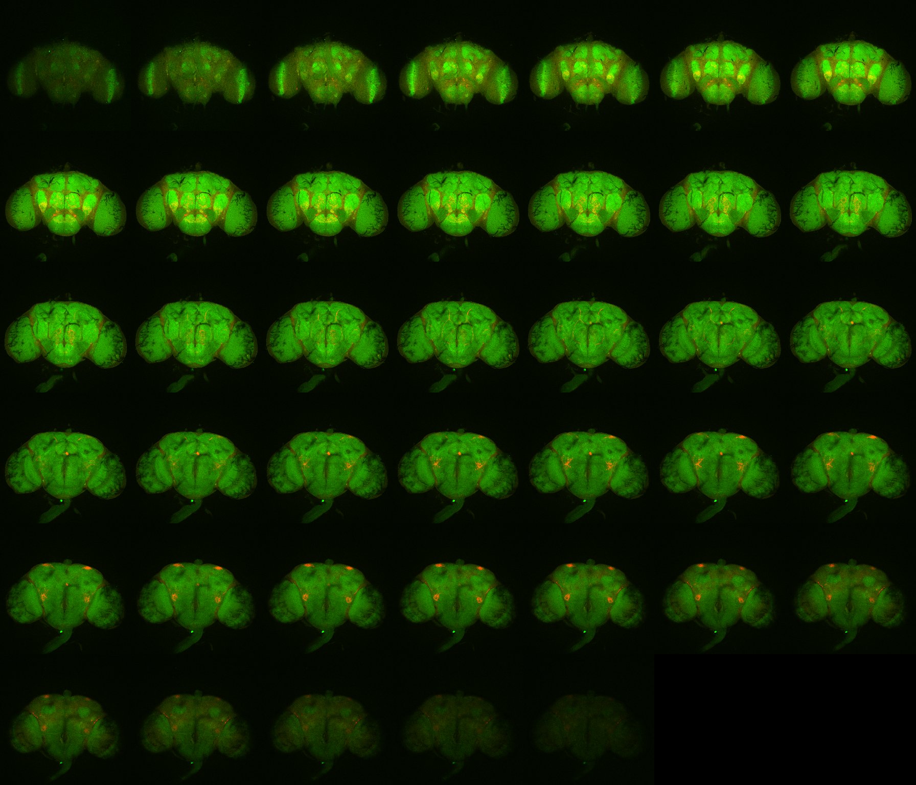
Image is a coronal projection of a confocal stack of a singular neuron type (FB2A) in the full volume of the Drosophila melanogaster brain. GFP staining should be in green and neuropil staining should be in magenta.
By Emily Kellogg, Rachel Wilson Lab, HMS
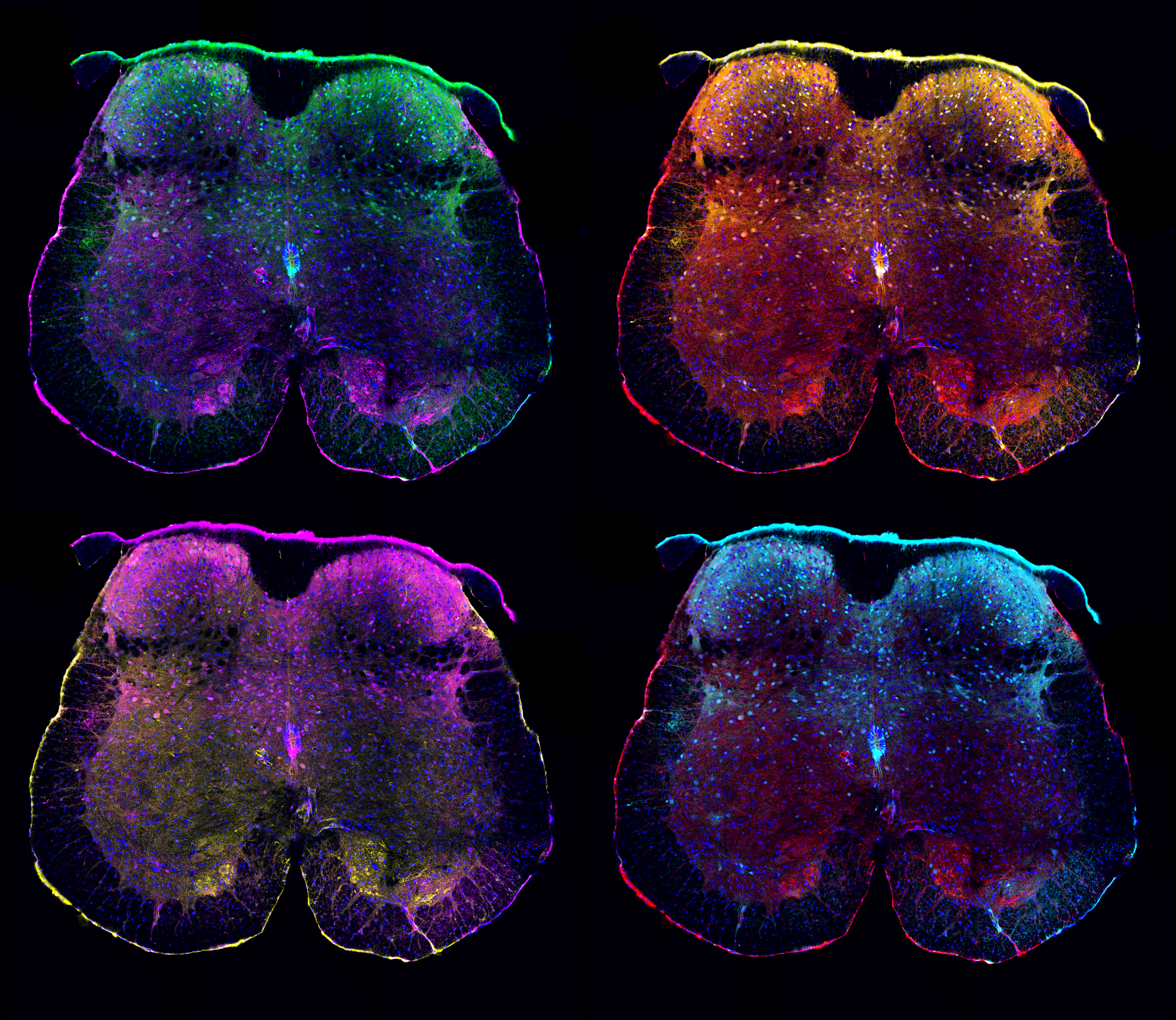
Leica SP8X
Sacral spinal cord neurons that express estrogen receptor 1 alpha (Esr1) were visualized using a transgenic reporter allele that encodes a red fluorescent protein, tdTomato. Sacral motor neurons were labeled using immunohistochemistry against choline acetyltransferase (ChAT). (Top left image – blue: cell nuclei, green: Esr1+ cells, magenta: ChAT+ cells)
By Trent Pottala, Sabatini Lab, HMS
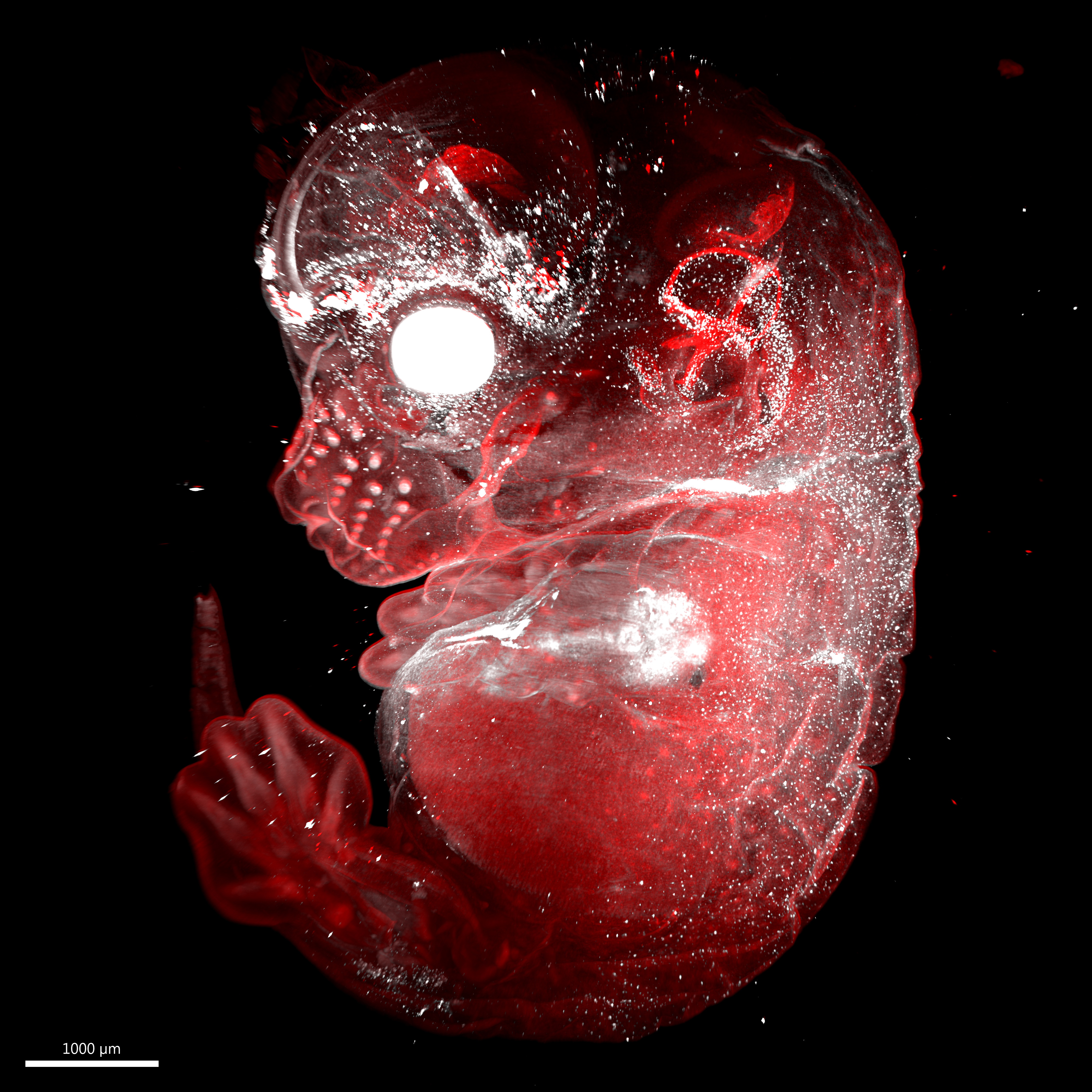
LaVision Lightsheet Microscope
Melanoblast cells migrating through the skin from the neural crest in an e13.5 embryo
By Brian Raftrey, Hsu Lab
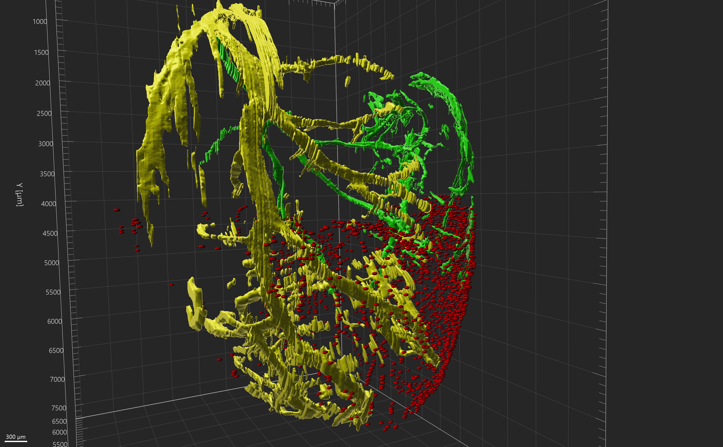
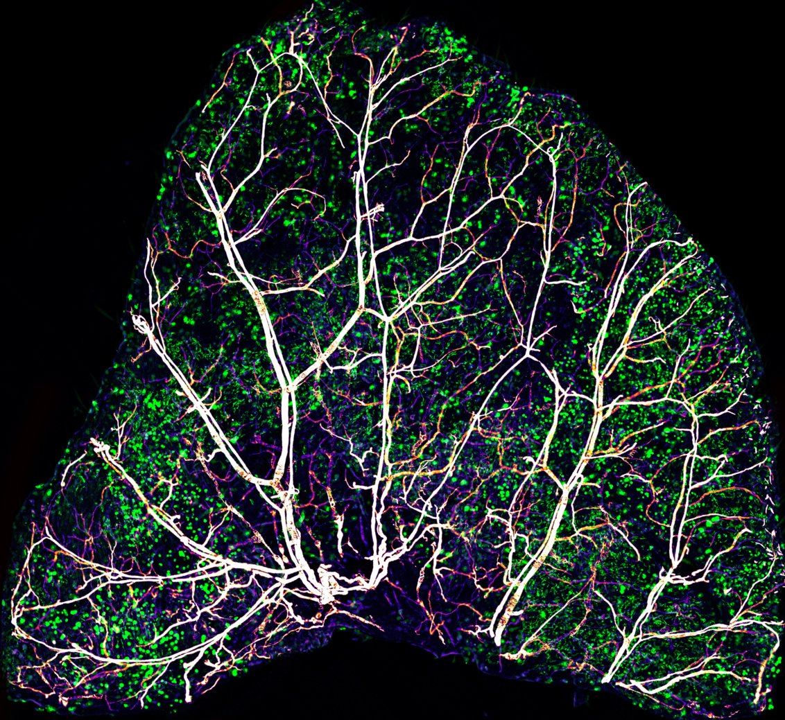
LaVision Lightsheet Microscope
Mouse heart cleared using iDISCO showing the left coronary artery (yellow), right coronary artery (green), and macrophages (anti-F4/80 - red dots) within a region of interest.
By Anurag Jamaiyar, Mark Feinberg Lab