Equipment


Coming Soon: The newest in slidescanning! The NIF has purchased 2 new VS200 whole slide scanners. One will have a 210 slide loader and one will have a 60x oil with oil dispenser. Expect delivery in early December.


List of available equipment


Neurolucida: Automated Neuron Reconstruction

Here's what they say about our software packages

https://www.mbfbioscience.com/products/microlucida 

Microlucida gives you the power to capture 2D and 3D whole slide images at your brightfield or fluorescence microscope. It allows you to easily generate high quality whole slide images and image stacks. It also enables you to use a single high-quality image acquisition application across all of your microscopes, a feature particularly beneficial for core facilities

**NEW**Leica Stellaris Confocal Microscope

The Stellaris is a fully spectral collection system with motorized stage for multi-point tiling. Excitation laser lines are 405nm-790nm. Objectives: 5x air 0.3NA, 20x multi-imme 0.75NA, 40x oil 1.3NA, 60x oil 1.40NA, 100x oil 1.44NA. 

The confocal has a Tokai Hit environmental chamber (heat, humidity and CO2 control) as well as silicone lenses (60x NA1.3, 100x NA1.35) for long time series experiments. 

Zeiss PALM Combi Laser Capture/Microtweezer Microscope

PALM CombiSystem combines two non-contact laser technologies in one system. It allows you to precisely microdissect and capture samples of interest and to optically trap small objects, such as living cells. PALM CombiSystem combines laser microdissection with optical trapping. For laser microdissection mark an element on your screen and then start the laser to separate it from unwanted tissue. A targeted laser pulse will ‘catapult’ your element upwards and contact-free into a collection vessel for further analysis.
The optical trap of PALM CombiSystem manipulates particles with the highest precision at the cellular and sub-cellular level. This allows you to trap, move and position microscopic objects — such as red blood cells and bacteria — intuitively with the force of light.


Whole Slide Scanning Microscopes

The facility houses 2 Olympus VS120 slide scanners that has been specifically designed for high throughput facilities that require fast scanning speed and high image resolution. The VS120-SL 5 Slide System holds 5 slides at a time making it quicker and easier to archive slides and produce permanent records. Imager can scan in 10x, 20x, and 40x. The systems have 2 cameras to collect Histolo or Immuno staining. Both systems have an Orca R2 monochrome (16 bit) camera for fluorescence (DAPI, Alexa 488, Alexa 560, Alexa 640 and Venus) and the same color camera for histology sections. 

LaVision LightSheet Microscope

The bidirectional triple light sheet technology of the UltraMicroscope II generates 6 focused light sheets to excite samples from the side while the fluorescence light is detected by a sCMOS camera perpendicular to the illumination plane. Moving the sample through the light sheet generates a 3D image stack. Selective excitation of the focal plane reduces bleaching and photo toxicity significantly. The open setup allows the analysis of cleared samples in any clearing solution or in aqueous media with and optical zoom from 1.26x to 12.6x . The system can handle samples up to 30 mm x 30 mm x 15 mm in size.


Lightsheet imaging can take all types of mounting media. Please inquire with us about what is the best for you


Widefield Microscope

The Olympus BX63  automated microscope is designed for brightfield, DIC and fluorescence staining. The system holds a 2x, 10x, 30x, 40x, and 60x objective with DAPI, GFP, TdTom, Cy5.5 filter cubes. This system can capture multiple channels of flourescence and can tile. The sample holder can hold 2 slides at a time for more efficient workflow.

Macro Scope

The MVX10 Marco dissecting microscope with fluorescence and brightfield capabilities with a high NA for collecting high resolution images of weak signal. This system is equipped with an Olympus DP80 camera that has the ability to switch between RGB color and monochorme imaging. The images are captured using CellSens software. CellSens has the ability to manually stitch large sections. Magnification of 0.63x -126x.

Leica SPE Confocal Microscope

The Leica TCS SPE is a prism-based spectral detection system. It has a highly dynamic PMT that offers extraordinary signal efficiency for gapless detection of weak signals. A variety of robust solid-state lasers allow the use of a broad range of common dye.

Available laser lines: 405nm (25mW), 470nm-670nm

Available Objective: APO 10x 0.30NA, APO 20x 0.60NA , APO 40x 1.15NA Oil, APO 63x 1.30NA Oil

  • Navigator function that allows for stitching tile scans
  • Spectral un-mixing of emission light
Leica SP8X STED Microscope

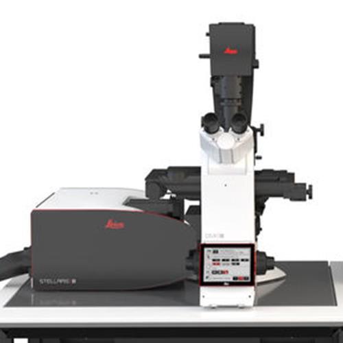
The Leica 3x STED/FLIM system is a super resolution microscope based on the Leica SP8 confocal microscope. It has a guaranteed resolution of 100 nm though the system has been routinely achieving 50 nm. We recommend referencing the accepted dye list and sample prep notes by Leica (https://www.leica-microsystems.com/science-lab/the-guide-to-sted-sample-preparation/) for STED.

The Fluorescent Life Time (FLIM) imaging has the ability to measure the decay rate of your probes. As the fluorescence lifetime does not depend on concentration, absorption by the sample, sample thickness, photo-bleaching and/or excitation intensity it is more robust than intensity based methods. 

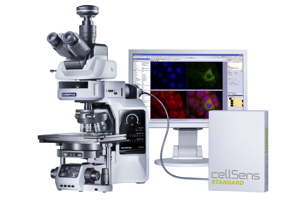
The confocal is equipped with multiple excitation lasers including a continuum fiber laser that can deliver up to 8 simultaneous lines of any wavelengths between 470 and 670 nm. It has 4 detectors, two of which are high-sensitivity Hybrid detectors. It can be operated in STED, FLIM or standard confocal mode. Feature highlights include:

  • White light laser: continuous excitation  tuning from 470 nm to 670 nm, Argon Ion Laser with 457, 477, 488 and 514 nm lines, 405 nm DPSS laser
  • 594nm, 660nm and 770nm Depletion laser
  • 4 HyD and 3 PMT detectors
  • Transmitted light detector
  • Objective lenses:10x dry, 20x dry, 40x oil, 63x oil for confocal, 100x oil.
  • Tokai hit incubation chamber
  • Navigation for tilescans
  • Laser-based autofocus
TissueCyte 2Photon Tomography

Imagine an imaging system that can image tile an entire organ using dual MPE excitation up to 1mm in depth then slices your sample at your pre-determined depth and image again. Your slices are automatically mounted onto a slide for later analysis.

You receive a high resolution 3D image of your entire organ with all your samples mounted on slides.

Simply drop off your samples and we do the rest. We will schedule your pickup of slides and data when it's finished. We are asking external users of this system to bring a hard drive (2TB+ depending on objective magnification).

https://www.tissuevision.com/